AI tools achieve high precision in cervical cancer staging and survival prediction
The study found that deep learning models, especially CNNs, were the most frequently implemented technique (61.2%), followed by traditional machine learning (34.2%) and hybrid ML–DL systems (4.6%). CNNs dominated in all three predictive tasks: classification, segmentation, and regression. SVM, random forest, and ResNet models were also widely used, while reinforcement learning and hybrid approaches made limited but promising appearances.
A review published in the journal Diagnostics provides a detailed analysis of how artificial intelligence technologies are transforming the diagnosis, prognosis, and treatment of cervical cancer - the fourth most common cancer among women worldwide.
The study titled “Clinical Applications of Machine and Deep Learning in Cervical Cancer: A Systematic Review” examined 153 peer-reviewed articles from 2019 to 2024, highlighting the predominance of deep learning models, especially convolutional neural networks (CNNs), and their significant potential in improving clinical workflows across multiple stages of cancer care.
How are machine learning models being used to diagnose cervical cancer?
Diagnosis is the most explored clinical application, accounting for 54.9% of the reviewed studies. Within this category, six primary diagnostic targets were identified: determining the stage of cervical cancer, conducting cancer screening, identifying recurrence, detecting HPV types, predicting cancer progression, and segmenting anatomical structures on medical images. The largest share of studies focused on identifying cancer stages and performing routine screening, often using colposcopy and cytology images as inputs.
Classification tasks were the most commonly employed method across all AI applications in cervical cancer, featured in over 70% of the studies. CNNs were the dominant model type for classification, chosen for their ability to extract visual patterns from medical imagery. These models demonstrated strong performance, with many achieving accuracy rates above 90%, validating their potential for clinical use. CNNs were particularly effective when analyzing cytopathology images, where precise cell shape and texture analysis are essential.
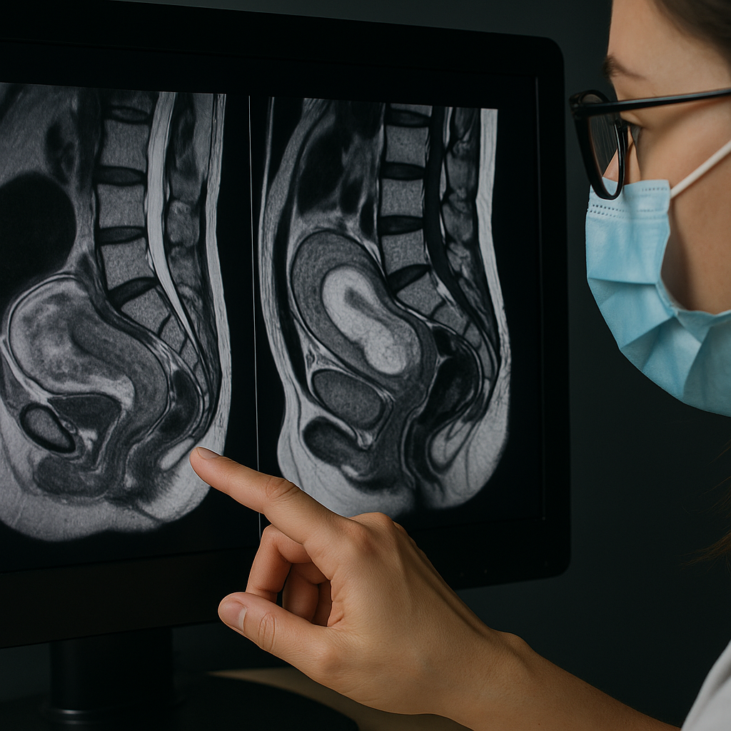
Several studies also implemented deep learning models to segment tumors and organs at risk (OARs) from MRI and CT images. These segmentation models were designed to support radiation therapy planning and achieve high Dice Similarity Coefficient (DSC) scores, reflecting their reliability in mapping out critical anatomical zones for targeted interventions. Furthermore, the review highlighted growing adoption of hybrid methods that combine both deep and traditional machine learning algorithms, yielding even higher diagnostic accuracy and robustness.
What role does AI play in prognosis and treatment planning?
Prognostic applications made up 22.9% of the studies and focused on three main targets: predicting cancer progression, estimating survival rates, and identifying recurrence risk. Many of these studies utilized multimodal data inputs, ranging from MRI radiomics to clinical history, to train classification and regression models. Machine learning models, particularly support vector machines (SVM) and random forest algorithms, were often employed to assess progression risks and survival outcomes, achieving accuracies around 85–90%.
Treatment-focused studies (22.2% of the total) emphasized automatic dose planning, target delineation, and segmentation for radiation therapy. The review revealed that U-Net and other CNN variants were commonly used to map clinical target volumes (CTVs) and estimate therapeutic doses. The highest-performing models reduced the need for manual intervention and showed accuracy levels competitive with expert-designed treatment plans.
Beyond dose planning, AI models were also tested for predicting radiotherapy toxicity, though such applications remained limited. Importantly, image-based treatment models were often validated using internal metrics but lacked external clinical testing, highlighting the gap between model development and clinical deployment.
Which AI techniques dominate and how are they validated?
The study found that deep learning models, especially CNNs, were the most frequently implemented technique (61.2%), followed by traditional machine learning (34.2%) and hybrid ML–DL systems (4.6%). CNNs dominated in all three predictive tasks: classification, segmentation, and regression. SVM, random forest, and ResNet models were also widely used, while reinforcement learning and hybrid approaches made limited but promising appearances.
In classification, CNNs were used in 40 studies, followed by SVM (25 studies) and random forest (12 studies). For image segmentation, CNNs led with 28 implementations, while regression tasks saw six CNN-based models for predicting radiation doses. These findings emphasize the versatility and adaptability of CNNs across various cervical cancer prediction challenges.
The review also analyzed validation practices. While 5-fold and 10-fold cross-validation were common, 33.96% of studies failed to report any validation strategy. Only 9.43% of machine learning studies conducted external validation, raising concerns about reproducibility and real-world readiness. This highlights the necessity for more rigorous testing and standardized reporting protocols in future research to ensure AI tools are clinically trustworthy.
- FIRST PUBLISHED IN:
- Devdiscourse


